Describe Anthony's Capsule Stain When Would You Use It
Capsules are formed by organisms such as Klebsiella pneumoniae. The Congo Red Capsule stain is a modification of the nigrosin negative stain you may have done previously.
Capsule Stain Principle Procedure Results Microbe Online
The positive capsule staining method Anthony Method uses two reagents to stain the capsular material.

. The capsule differs from the slime layer that most bacterial cells produce in that it is a thick detectable discrete layer outside the. Anthonys method uses Crystal violet 7min then rinses with 20CuSO4. INTRODUCTION As mentioned in lab exercise 6 not all dyes stain the bacterial cell.
DrWhitneyHolden goes over bacterial capsules - what a capsule is and two different methods for how to stain it. In both Klebsiella and Bacillus capsule can be seen surrounding the dark purple coloured cells. The capsule or glycocalyx is a gelatinous outer layer that is secreted by the microbe and remains stuck to it.
The waxy acid-fast cells retain the. Below describe these characteristics for both bacteria. When you interpret a Gram stained smear you should also describe the morphology shape of the cells and their arrangement.
It divides most of the EUBACTERIA into two large groups. The bacteria take up the congo red dye and the background is stained then with acid fuchsin dye. It is widely used in the microbiology laboratory for the staining of.
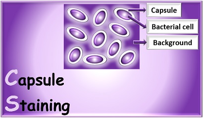
Cells lacking capsules will have a gray-ish stain from the Congo red dye directly adjacent to the pink colored cell body. The principle of capsule staining is based on staining of background with an acidic stain and staining of bacterial cell with a basic stain. In Figure 5 there are two distinct types of bacteria distinguishable by Gram stain reaction and also by their shape and arrangement.
June 5 2021 by Sagar Aryal. This is a DIFFERENTIAL STAIN. The basic procedure goes like this.
Capsules may be polysaccharide glycoproteins or polypeptides depedning on the organism. List two functions of a capsule. The capsule stain employs an acidic stain and a basic stain to detect capsule production.
The primary stain Crystal violet is applied over a non heat fixed bacterial smear so that both the bacterial cells and capsular material take up the color of the primary stain. Perform a negative staining procedure 2. This one is another most commonly used method of Capsule staining.
Flood the smear with CRYSTAL VIOLET 1 minute then wash with water. Perform a successful capsule stain to distinguish capsular material from the bacterial cell. Congo Red Capsule Stain.
Cells that have a heavy capsule are generally virulent and capable of producing disease since the structure protects. Identify the name and type of dye used to stain the cells in a capsule stain. Thus capsule staining creates contrast by staining a bacterial cell along with its background and leaving a capsule as a colourless halo.
The Capsule of the bacterial cell appears as a clear halo around the stained bacterial cell body and the Dark background color depends upon the dye used India ink or Nigrosin. Those that have waxy mycolic acids in their cell walls and those that do not. Stain Protocols BIOL 2420 Smith 2010 Page 3 of 4 Capsule Stain 1.
An acid-fast stain is able to differentiate two types of gram-positive cells. Pneumoniae or the organism you want to stain from your tube or plate and mix it into the drop of India ink. Capsule will appear as a clear zone in negative staining against dark background whereas it is light blue coloured in.
Giemsa stain is a Romanowsky stain. A capsule being non-ionic will not stain by either of the two dyes. It requires a PRIMARY STAIN and a COUNTERSTAIN.
After the primary stain is applied and the smear is heated both the vegetative cell and spore will appear green. Place a single drop of India ink on a clean microscope slide adjacent to the frosted adge. It helps bacteria attach to the host tissue prevents the cell from drying out.
The capsule stain employs an acidic stain and a basic stain to detect capsule production. CAPSULE STAIN Antonys MethodPrinciple. The other approach of capsule staining.
Understand the benefits of using negative stains. I also have vi. For further penetration the application of heat is required.
Describe the visual appearance of stained cells that would allow you to determine if a cell lacks a capsule. Take a heat fixed bacterial smear. Both use carbolfuchsin as the primary stain.
Apply dye stain crystal violet or methylene blue for 1-2 min then rinse. Apply oil and look at the shapes arrangements etc. Two different methods for acid-fast staining are the Ziehl-Neelsen technique and the Kinyoun technique.
Capsule Stain- Principle Procedure and Result Interpretation. It is not common to all organisms. Heat fix by passing slide through the flame on a clothes pin 3x to kill bacteria make them stick 5.
The only explanation I have found for this method states that the crystal violet stains the cell and the capsule purple and the CUSO4 decolorizes and counterstains the capsule light blue. It takes out the colour from the capsular material and counter stains the decolourized capsule which now appears as a light blue zone surrounding deep purple cells. Principle of Capsule Staining Capsules stain very poorly with reagents used in simple staining and a capsule stain can be depending on the method a misnomer because the capsule may or may not be stained.
It helps to demonstrate the presence of capsule in bacteria or yeasts. Streptococcus pneumoniae Neisseria meningitidis Haemophilus influenzae Klebsiella pneumoniae are common capsulated bacteria. The Negative Stain and Capsule Stain OBJECTIVES 1.
Stick it between blot paper. In this technique the crystal violet stain is used as the primary stain. The capsule or slime layers highly hydrated polymers exclude both dyes.
Both the cell and the capsule become stained by the crystal violet. Most capsules are composed of polysaccharides but some are composed of polypeptides. Unlike most vegetative cell types that stain by common procedures the free spore because of its impervious coats will not accept the primary stain easily.
Using a flamed loop and sterile technique remove some K. GRAM POSITIVE bacteria and GRAM NEGATIVE bacteria. The capsule will appear as a white halo between the red cell and the darker background.
Principle of Spore Stain. A capsule is a gelatinous outer layer that is secreted by the cell and that surrounds and adheres to the cell wall.
Characterization Of D Cpxar Mutant A Anthony S Capsule Staining Was Download Scientific Diagram
Capsule Staining Principle Methods Procedure Result Interpretation
Capsule Staining Procedure Anthony S Stain Method Part 7 Youtube
Capsule Staining Principle Reagents Procedure And Result Online Biology Notes
Anthonys Staining Method For Capsule Staining Ll Hindi Tutorial Ll Biology Youtube
Capsule Stain Principle Procedure Results Microbe Online
Capsule Staining Principle Methods Procedure Result Interpretation
Capsule Stain Principle Procedure And Result Interpretation
Capsule Staining Principle Methods Procedure Result Interpretation
What Is Capsule Staining Definition Principle Methods Biology Reader
Capsule Staining Principle Methods Procedure Result Interpretation
Capsule Stain Definitions Methods And Procedures
Capsule Staining Principle Reagents Procedure And Result
Endospore Staining V The Name Endospore Is Suggestive
Anthony S Capsule Staining Microbiology Note Facebook
What Is Capsule Staining Definition Principle Methods Biology Reader
Anthony S Capsule Staining Microbiology Note Facebook
Endospore Staining The Name Endospore Is Suggestive Ppt Download
Comments
Post a Comment